Flat Foot Correction:
FDL transfer and Calcaneal Osteotomy.
(In Posterior Tibial Tendon Dysfunction)
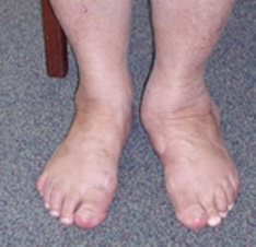

Posterior tibial tendon dysfunction is a very common problem. It occurs when the posterior tibial tendon becomes inflamed or torn. As a result, the tendon may not be able to provide stability and support for the arch of the foot, resulting in flatfoot. This operation is for ankle pains due to flat feet (pes planovalgus). The pain is due to inflammation of the posterior tibial tendon, this muscle / tendon supports the arch of the foot. The inflammation allows the tendon to stretch or tear, causing or exacerbating a flat foot deformity. As the deformity progresses, it causes arthritis of the hindfoot joints and pain at the outside of the ankle. This type of operation is used before the deformity has progressed to arthritis.
The surgery involves a transfer of the flexor digitorum longus tendon (FDL) into the navicular bone, to take the place of the diseased tendon. The operation is performed through an incision running from the inside ankle to the arch of the foot. It is combined with an osteotomy (cut) of the Os Calcis (heel bone) to reinforce the deformity correction. The osteotomy is performed through an incision on the outside of the heel and fixed with a screw.
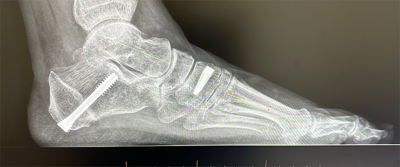 Restoration of the arch of foot and Calcaneal screw
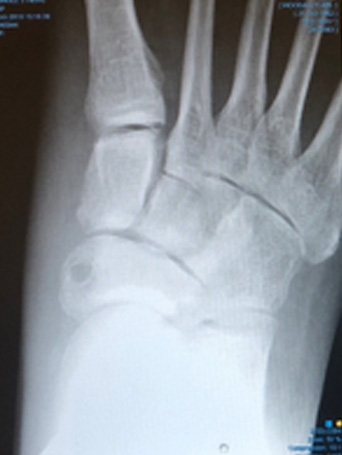 Hole where FDL tendon transfer passes through navicular bone
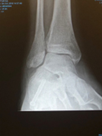 Screw fixing the calcaneal osteotomy / heel shift.
Preparation for Surgery
You should have received a letter detailing the codes and costs for the surgery and giving instructions on where to go and at what time. Please check these details carefully as you will be liable for any costs not covered by your insurers.
You will need to be nil-by-mouth – No food for 6 hours before surgery. Clear fluids can be taken for up to 2 hours before the operation.
This operation normally requires an overnight stay of 1-2 days.
MRSA status
Before or on admission to hospital a nasal swab will be taken to screen for MRSA. There is a small chance this is positive. If so your operation will be moved to the end of the list or rarely to another day to prevent cross infection.
Anticoagulants
If you are taking an anticoagulant, this will need to be stopped / changed prior to the surgery. Please discuss this with the specialist that prescribed it. Your surgery cannot go ahead without this.
Anaesthetic
The surgery is performed under a general anaesthetic. On top of this you will be given a regional block - an epidural of the leg which numbs the leg for 1-3 days, providing excellent pain relief.
Immediately after surgery
After surgery, your leg will be immobilised in a backslab (half plaster) for 2 weeks. Elevation of the foot (above the pelvis) for the first 2 weeks is vitally important to prevent infection. Naturally, small periods of walking and standing are necessary, but no weight must be taken through this leg for 6 weeks.
You will be seen by a physiotherapist on the ward, who will advise on using crutches and your rehabilitation. You will be allowed home only when you are comfortable and capable.
Weight-bearing
You cannot walk on the foot for 6 weeks. Initially you will be an plaster cast for 2 weeks and then a protective boot for 4 weeks. Walking can start at the 6 week stage, in the protective boot + an insole. It will take another 4-6 weeks to be able to walk without the boot and several months to walk normally.
Using crutches can be difficult even for the able-bodied. There are some useful aids that can be bought or hired.
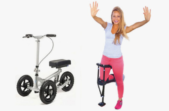
The knee rover is a scooter with brakes that takes the weight of the leg. www.kneerover.com
The iWalk is a ‘peg-leg’ – it requires good balance. www.peglegs.co.uk
Risks of surgery
Swelling
Initially the foot will be very swollen and needs elevating. The swelling will disperse over the following weeks & months but will still be apparent at 9-12 months.
Infection
This is the biggest risk with this type of surgery. You will be given intravenous antibiotics to prevent against it. The best way to reduce your chances of acquiring an infection is to keep the foot elevated for 2 weeks. If there is an infection, it should resolve with a course of oral antibiotics. Very rarely a severe infection occurs, requiring IV antibiotics as an inpatient.
Nerve Damage
Alongside the incisions are two nerves – the saphenous and the sural nerves. They supply sensation to the sides of the foot and toes. They may become damaged during the surgery and this will leave a patch of numbness at the sides of the foot. This numbness may be temporary or permanent. There is approximately a 5% of this happening.
Failure to relieve all symptoms
The results from this surgery are good. Published studies show that over 90% of people have good relief of their pain for more than 10 years.
Recurrence
In 50%, there is some recurrence of the deformity, but without pain. It is very unusual to need further surgery following this procedure.
Blood Clots
There is a small risk of developing a DVT and to help prevent this you will be prescribed a daily anticoagulant to take until walking.
Recovery from surgery
Please make an appointment for a review at 2 weeks, when the backslab will be removed and the stitches taken out. The plaster cast is changed for a protective boot at that stage and physiotherapy can start. You will be reviewed again 4 weeks later, when you can start walking in the protective boot with an arch support.
You will be reviewed again at 3 months following surgery, with x-rays.
Physiotherapy is an essential part of your recovery and will start at 2 weeks. Naturally you will get stronger each week but it will take many months to regain full activity.
Pain relief and take home medications
You will be given high doses of prescription painkillers to take home. Use these for the first 3-4 days and reassess.
Washing and Bathing
It’s important to keep the dressing completely dry – the nurses will show you how to do this with a waterproof cover.
Activity and time off work
In general, up to 4 weeks off work is required for sedentary posts. 12 weeks for standing or walking posts. 16 weeks for manual / labour intensive posts.
When can I start to drive again?
The DVLA states that it’s the responsibility of the driver to ensure they are always in control of the vehicle. With surgery to the left leg, driving an automatic is permitted after 2 weeks. On the right leg, driving will not be possible until 6-8 weeks.
It remains your responsibility to drive safely and you should also check with your vehicle insurer to confirm you are covered.
Results of surgery
The short to mid-term results of this type of surgery are very good and the majority of patients get excellent relief for many years. It may be better to consider this as an interim procedure, as the underlying arthritis will continue to affect the joint and so symptoms can often recur after several years.
Revision 18.05.2019