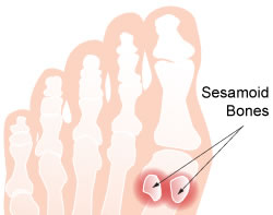
Sesamoid Bones
What are they?
The two semsamoid bones (medial and lateral) are two 1 x 1.5cm oval shaped bones that lie under the big toe joint, within the two tendons that move the big toe (flexor hallucis brevis). They act in a manner similar to that of the knee cap, acting to straighten the big toe. To achieve this functionality, the semamoid bones have to be very smooth, to allow them to glide smoothly over the head of the first metatarsal bone, and to this end they are covered with a smooth layer of articular cartilage.

How do they get damaged?
The sesamoid bones are routinely put under a lot of stresses, as they transmit a great deal of the force on the foot in the act of walking. Repetitive injury may damage the articular cartilage, roughening its surface, leading to painful inflammation. In some cases the bones may also fracture. This is an injury directly related to over use, and most commonly occurs in sportsmen such as sprinters & jumpers.
The primary symptom is pain felt under the big toe joint, and may be constant and acute, causing a limp. In some cases chronic pain may be felt sporadically by sportsmen, who have no symptoms during normal activity, but aggravate the injury when performing sporting activities.
Diagnosis
The foot is first carefully examined to exactly pin point the location of the pain, and to exclude other possible causes of big toe pain. This examination is then followed up with X-rays or MRI scanning to ascertain the cause and extent of the injury.
Treatment
The first step in treatment is non-surgical, and requires a change in the exercise regimen, designed around reducing the stress on the sesamoid bone to allow recovery, but with still enough activity to prevent the stiffening of the toe joint. To this end a special shoe insert may be employed, designed to distribute the forces experience by the big toe across the entire foot. Once the initial acute episode has settled, a progressive return to former activity levels may commence.
In most cases, this program of rehabilitation is sufficient to alleviate the problem, but for those patients who fail to see results with the above techniques, surgery is an option. In the case of a fracture a one graft, or repair with a tiny screw may be performed. In severe cases the affected sesamoid bone may be removed. If removal of the sesamoid is necessary, minor surgery is also performed to repair the flexor hallucis brevis tendon, to ensure that good function and push of strength is retained.
Prognosis
Most patients are able to return to previous activity levels after their rehabilitation is complete, and even those who require surgery are generally able to return to full strength. The time for rehabilitation will depend on the severity of the initial injuries, but most should see a full recovery within a few weeks.