Achilles Repair and Rehabilitation

The goal of treatment is to have an Achilles and calf that functions as best as possible, allowing full activity and sport.
The Achilles tendon is exceptionally strong and, as yet, we don’t know why it ruptures. It often happens during the simplest activities and you and any spectators may have heard it snap. Patients often say it feels as though they were hit from behind.
When the tendon ruptures, It is very much like a rope snapping with two frayed ends. The tendon heals with scar tissue and it’s very important that they are as close together as possible. They should then heal with the optimum tension giving the muscle maximum power once recovered. If the tendon ends gap then this leaves the muscle lax and weaker.
Naturally, because the Achilles is so strong, it takes a long to time to heal fully. It will take several months to be functioning well and over a year to heal fully. Research has shown that a tendon heals best if it is carefully moved in a protected environment – therefore Physiotherapy is an essential part of this and you will get to know your physio well!
Preparation for Surgery
You should have received a letter detailing the codes and costs for the surgery and giving instructions on where to go and at what time. Please check these details carefully as you will be liable for any costs not covered by your insurers.
You will need to be nil-by-mouth – No food for 6 hours before surgery. Clear fluids can be taken for up to 2 hours before the operation.
MRSA status
Before or on admission to hospital a nasal swab will be taken to screen for MRSA. There is a small chance this is positive. If so your operation will be moved to the end of the list or rarely to another day to prevent cross infection.
Anaesthetic
The surgery is normally a daycase procedure, performed under a general anaesthetic with local anaesthetic after for additional pain relief. It is not normally a particularly painful operation and most patients report minimal pain.
Operation
The Achilles is repaired using a minimally invasive (MI) or keyhole technique. This is performed through a 1-2cm transverse incision over the rupture site. The keyhole surgery causes minimal trauma to the Achilles sheath and skin and so has the lowest risks. It also allows rapid healing with benefits in early rehabilitation. The surgery is performed with strong permanent sutures passed using MI methods. Even though the incision is small, the tendon repair can be assessed and visualised to ensure that the best tension and repair has been achieved.
Immobilisation
At the end of the surgery, the leg is immobilised in a plastercast with a soft bandage at the front or “backslab”, to allow for any swelling. You will be discharged in this, on crutches non-weight bearing (NWB). The cast will be changed at 10-14 days to a protective boot for 8-10 weeks.
Risks of Surgery
It is important to be aware of the risks of surgery. The vast majority of patients have no problems, but complications can happen and we do our best to minimise these risks.
Infection
Approx 1% risk. Usually this is a small infection of the skin that can be treated with oral antibiotics. Very rarely, a deep infection occurs requiring further surgery and this will compromise the final outcome.
Nerve Injury
Risk 1%. The sural nerve is a sensory that supplies sensation to the skin of the side of the foot and heel. It can be temporarily or permanently damaged. Usually no further treatment is needed. Rarely the nerve will be painful requiring further surgery.
Re-rupture
Risk 2-3%. This is usually due to a slip or fall.
Blood Clots
Risk 2-5%. You will be given an oral anticoagulant to protect against this. If it does occur, this is treated with oral anticoagulants for 3 months.
Immediately after surgery
You will be referred to a physiotherapist who will advise on walking while keeping weight off the ankle using crutches. You will be sent home only when you are comfortable.
Pain relief and take home medications
You will be given high doses of prescription painkillers to take home. Use these for the first 2-3 days and reassess.
There is a small risk of blood clots “DVT’s” with Achilles surgery and you will be prescribed a blood thinner for 2 weeks.
Weight-bearing
Initially you will be Non WB for 2 weeks then partially WB in a protective boot, with Full WB at the 4 week stage.
Using crutches can be difficult even for the able-bodied. There are some useful aids that can be bought or hired.
-
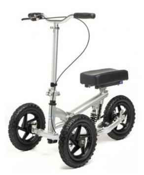 The knee rover is a scooter with brakes that takes the weight of the leg. www.kneerover.com
 The iWalk is a ‘peg-leg’ – it requires good balance. www.peglegs.co.uk
Rehabilitation
Vacoped Rehab Achilles Rupture
| WIEGHTBEARING PHASES | |
| 0-2 weeks Non-Weight Bearing (NWB) in POP | |
| 2-4 weeks Partial Weight Bearing (PWB) in boot up to 50% | |
| 4+ weeks Full weight bearing (FWB) in boot | |
| 10+ Weeks FWB in Shoes | |
| Phase 1 | Protection (0-8 weeks) |
| Phase 2 | Early Mobilisation (6-12 weeks) |
| Phase 3 | Strengthening (12-20 weeks) |
| Phase 4 | Return to Activity (5-7 months) |
| Phase 5 | Return to play (8-12 months) |
| WEEK 1 & 2: SWELLING MANAGEMENT Patient guide
Physiotherapy guide
|
|
WEEK 3 & 4: EARLY REHAB & INCREASING RANGE OF MOTION Patient guide
Physiotherapy guide
Physiotherapy guide
|
| WEEK 5 & 6: FULLY WEIGHT BEARING (FWB) IN BOOT & PROGRESSIVE WB AND REHAB Patient guide
Physiotherapy guide
|
| WEEK 7 & 8: GRADUAL INCREASE IN ACTIVITY Patient guide
Physiotherapy guide
|
| WEEK 9: LIGHT NON-WIGHTBEARING SPORTS Patient guide
Physiotherapy guide
|
| WEEK 10-13: FWB in shoe Patient guide
Physiotherapy guide
|
| WEEKS 13-20: Progress to Full Sports Patient guide
Physiotherapy guide
|
Milestones – Rehab goals
WEEK 16 onwards to RTS
|
WEEK 20 onwards
|
FAQs
When can I drive?
Manual - When you can do an emergency stop safely no earlier than 8 weeks
Automatic with left leg week 2
When can I return to work?
Sedentary – 2 weeks WFH and week 4 return to office
Manual labour – 12-16 weeks return to work
When can I cycle outdoors?
16 weeks post op can safely bike outdoors
When can I jog?
20 weeks post op with supervision of this milestone with Physio
When can I swim?
With a Vacoped boot start hydro with supervision of Physio at week 4 onwards
Swim only weeks 16
When can I play tennis?
4-6 months post to discuss milestone with physio
When can I wear normal shoe?
Week 15 onwards
When can I get back on the pitch?
Elite athlete – 6-9 months
Recreational athlete – 9 months
When can I wear high heels?
20 weeks onwards
When can I ski?
6 months post discuss with physio and consultant
Operative treatment versus nonoperative treatment of Achilles tendon ruptures: systematic review and meta-analysis
BMJ 2019
Conclusions
This meta-analysis shows that operative treatment of Achilles tendon ruptures reduces the risk of re-rupture compared with nonoperative treatment. However, re-rupture rates are low and differences between treatment groups are small (risk difference 1.6%). Operative treatment results in a higher risk of other complications (risk difference 3.3%). The final decision on the management of acute Achilles tendon ruptures should be based on patient specific factors and shared decision making. This review emphasises the potential benefits of adding high quality observational studies in meta-analyses for the evaluation of objective outcome measures after surgical treatment.